

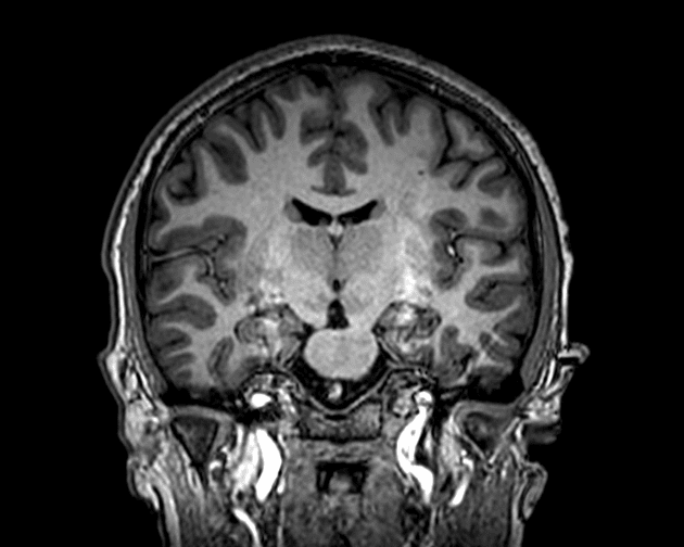
Introduction
Magnetic Resonance Imaging (MRI) is a powerful tool in modern medicine, but it is not without its challenges. One of the main challenges radiology students face is understanding MRI artifacts. These artifacts are distortions or errors in MRI images that can affect diagnosis if not recognized correctly. Knowing the common types of artifacts can help students and professionals produce more accurate images and improve patient care.
Understanding MRI artifacts is crucial because they often mimic or obscure real anatomical structures. Some artifacts are caused by patient movement, while others result from technical or hardware issues. Recognizing these patterns early in training can make a significant difference in interpretation and reporting accuracy.
Motion Artifacts
Motion artifacts occur when a patient moves during the scanning process. Even slight movements, such as breathing or swallowing, can blur the image. Motion artifacts often appear as ghosting or streaking, which can make it difficult to identify small lesions or subtle anatomical changes.
To reduce motion artifacts, patients should be instructed to remain as still as possible during the scan. In addition, radiology technicians can use faster imaging sequences or immobilization devices. Understanding this type of artifact is essential for radiology students, as it is one of the most common challenges encountered in daily MRI practice.
Metallic Artifacts
Metallic objects, such as dental fillings, implants, or surgical clips, can cause severe MRI artifacts. These artifacts appear as bright or dark areas on the image, often distorting the surrounding anatomy. Metallic artifacts occur due to the interaction of metal with the magnetic field, creating local field inhomogeneities.
Radiology students should learn to recognize metallic artifacts to avoid misdiagnosis. Patients are usually screened for metal before an MRI, but some implants are unavoidable. Techniques such as using different imaging sequences or specialized software can help reduce the impact of metallic artifacts on the final image.
Chemical Shift Artifacts
Chemical shift artifacts happen due to differences in resonance frequencies between fat and water molecules in the body. These artifacts typically appear as bright or dark lines at fat-water interfaces, often around organs like the liver or kidneys. Chemical shift artifacts are subtle but important to recognize in clinical imaging.
Awareness of chemical shift artifacts helps students distinguish between actual pathology and imaging errors. Adjusting imaging parameters or using fat suppression techniques can minimize these artifacts, leading to clearer and more reliable images.
Susceptibility Artifacts
Susceptibility artifacts occur when there are variations in the magnetic properties of tissues or foreign objects. These artifacts are common near areas like the sinuses, lungs, or metallic implants. On MRI images, susceptibility artifacts appear as distortions, signal loss, or bright spots.
Radiology students must understand susceptibility artifacts because they can mimic pathological findings such as hemorrhages or tumors. Recognizing and correcting for susceptibility artifacts allows for better diagnostic accuracy and reduces the risk of misinterpretation.
Wrap Up
Learning about MRI artifacts is essential for any radiology student. These artifacts, including motion, metallic, chemical shift, and susceptibility types, can significantly impact image quality and interpretation. By understanding the causes and appearances of these artifacts, students can improve their diagnostic skills and provide better patient care.
Recognizing MRI artifacts early in training helps radiology professionals avoid misdiagnosis and enhances the quality of imaging studies. With practice, students will become more confident in identifying artifacts and differentiating them from real pathology, making them more competent and reliable in their future careers.